
The completion of various preliminary studies on a work allows us to know all its characteristics: nature and quality of materials, unions and assemblies, as well as the state of conservation of different layers that make it up. This way, with the correct interpretation of the results obtained, we achieve a diagnosis of the work in accordance with its state of conservation, for an appropriate restoration and conservation treatment.
Such studies or preliminary analyses can be destructive, accelerate deterioration or natural aging of original materials, so it is vital to establish an order when conducting such preliminary examinations, always ensuring the safety and durability of the work.
Preliminary studies and examinations.
We can differentiate two types of examination methodology: using visible wavelength and using non-visible wavelength.
- Preliminary studies and examinations with visible wavelengths:
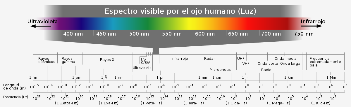
When we refer to visible wavelengths, light waves or the visible spectrum (fig. 1), we are talking about the range of the electromagnetic spectrum that the human eye is capable of perceiving. We call visible light or simply light this range of electromagnetic radiation with wavelengths that are visible to the human eye. Generally, we can capture wavelengths ranging from 380-400 nm (nanometers) which is blue color, to 700-780 nm which is red color (López and Vergara, 2017, p. 62)
Therefore, within that electromagnetic spectrum are the following preliminary examinations:
- Organoleptic examination with normal light: consists of examining the work using senses, especially sight. Additionally, by combining different positions of light points, much information about the work can be obtained, such as execution technique, deterioration or previous interventions.
- Normal light: The beam of light is directed directly onto the surface of the work. It is recommended that the light source used is LED type, which allows for a clearer and sharper observation of the work, as well as highlighting the natural brilliance of its colors (Muros and Revelo, 2018, p. 18). Among its advantages, we can highlight that they do not emit UV (ultraviolet) or IR (infrared) radiation, also representing an energy and economic saving (Silva, 2020: 15).
- Raking light: The beam of light is directed tangentially to the surface. This makes it possible to appreciate irregularities, textures, blisters, deformations, etc. (Cardell, 2008, p. 11).
- Transmitted light: the light is placed on the opposite side to what the observer sees, in this way, the light passes through a semi-opaque body.
- Organoleptic examination with ultraviolet light: consists of exposing the work in a dark space to ultraviolet radiation, which works with wavelengths less than 400 nm. As a result, materials that make up the work emit fluorescence that we perceive visually due to the excitation of ultraviolet light, allowing us to distinguish oxidized varnishes and locate previous interventions (repainting, additions) that are not perceptible to the naked eye under normal light. Prolonged use of these radiations can be harmful to health, so limited exposure is recommended.
For example, Guurtje Van de Stadt's portrait, by Claude Monet (fig. 2, left), has undergone conservation treatments mainly consisting of the removal of a yellowish and shiny varnish that hid the original cooler colors of the painting. This varnish reacts under UV light (fig. 2, right) and it is perfectly visible where this varnish was removed, namely in the lower left corner and upper right corner and on the face.

- Studies of microscopy and sample extraction: in this type of study, it is about knowing the different layers present in the work, their sequence, appearance, and state of conservation.
- Preliminary studies and examinations with non-visible wavelengths.
The invisible spectrum is the part of the electromagnetic spectrum that cannot be perceived by the human eye, this range of the electromagnetic spectrum is also called non-visible wavelengths.
Within the invisible spectrum, there are two regions: on one hand, infrared rays, which include infrared radiation, radio signals, television, microwaves and thermal radiation, working with a wavelength greater than 750 nm. On the other hand, ultraviolet rays, which include ultraviolet rays, X-rays and gamma rays, working with wavelengths less than 400 nm (fig. 1).
The study of works with non-visible wavelengths allows us to know data that are difficult to perceive with the naked eye about the work, especially when it comes to the internal structure of the work and its components.
- X-rays
It is a non-destructive analytical technique that facilitates the study of works, however:
"These studies are complex to interpret and provide limited information. Although X-ray studies allow us to perform an internal analysis of the work, this poses a series of inconveniences such as plane superposition (…) the information provided by the radiograph (…) although it is useful for polychromy studies, it is limited in terms of execution technique and conservation state." (Sarrió, 2015, p. 25)
As an example of this technique, the radiograph performed on Isabel Porcel's portrait, a painting by Francisco de Goya from around 1805, is shown revealing another woman underneath (fig. 3).

- Computerized Axial Tomography with X-rays (CAT)
It is a non-destructive analysis technique, more recent and advanced than radiographic studies, although not widely used. It allows obtaining and working with cross-sections and high-resolution 3D images without superposition, of millimeter thickness and the interior of any object that can be penetrated by an X-ray beam (Juanes, 2010, p. 32).
"These images have a high contrast that allows distinguishing between materials with a density difference less than 1%" (Sarrió, 2015, p. 28)
Conclusions
Preliminary studies, diagnostic techniques and analysis methods favor research on the work of art and provide significant data: dating, constituent materials, execution technique, state of conservation, etc. That allows us to justify, support and decide the best method, criterion or material to use regarding treatments to be carried out and what are the ideal conditions for conserving the work.
Despite CAT analysis providing the most information about internal structure, execution technique, and conservation state of the work, this type of study is economically inaccessible and its maintenance is costly. Additionally, it involves emitting X-rays, although not in large quantities, prolonged exposure to such radiation can have harmful effects on health.
For these reasons, the most commonly used techniques are those carried out with direct normal light, raking and transmitted light since they are non-damaging to the work or restorer, always using LED lighting.
On the other hand, UV light is mainly used to know the conservation state of the protective layer of the work and locate subsequent interventions that are not perceptible with the naked eye. In some cases, small samples are extracted to know stratigraphy and composition; these should be as minimal as possible and taken from representative areas that can provide the most data about original materials.
All preliminary studies, diagnostic techniques, and analysis methods must be carried out with the premise of maximum respect for the integrity of the work. The restorer is the professional best suited to decide which studies and analyses should be performed.
Bibliography
ABC.es. (04/07/2020). The Prado Museum Reveals the Secrets of Its Works Through X-rays. abc. Retrieved from: https://www.abc.es/cultura/abci-museo-prado-desnuda-algunas-40941447088-20200704012123_galeria.html [ 20/03/23]
Cardell Fernández, C. (2008). Visual examination and state of conservation of the internal architectural decoration of tomb QH33 in Qubett El-Hawa Aswan (Campaign 2008). Boletín de la Asociación Española de Egiptología, 18, 7-19.
Fernández Ruíz, E. (2008). X-ray of the Virgin of Blood. Instituto Andaluz del Patrimonio Histórico. http://hdl.handle.net/11532/16465
Juanes, D. (2010 ) Computerized Axial Tomography. Study of wood sculpture. In The Science and Art II: Experimental Sciences and Conservation of Historical Heritage (pp. 32-43). Ministry of Culture.
López Vergara, I & Vergara Alonso, M.H. (2017). Visible spectrum and digital images as a learning resource. In M. A. Cienfuegos Velasco et al (Eds.), DIALOGUES in Praxis: Views and knowledge of educational actors (pp. 61-74). Autonomous University of the State of Mexico.
Muros Alcojor, A., & Revelo Morales, N. L. (2018). Technical and perceptual comparison of LED and halogen lighting for a painting. icandela, (24), 12-18.
Silva Fino, M. (2020). A spectral analysis system optimizes the illumination of works of art and cultural heritage. PH: Bulletin of the Andalusian Institute for Historical Heritage, 28 (100), 15-17.


.jpg)
Comments